Human Nervous System
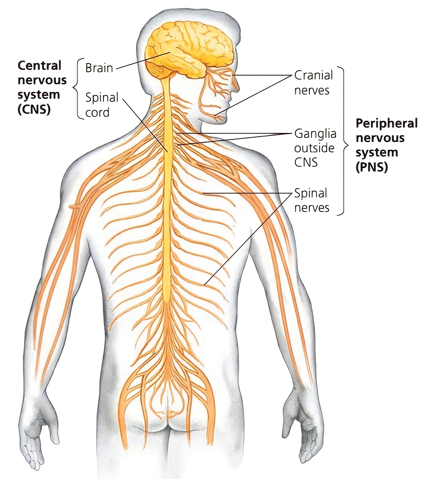
The nervous system is the part of an animal's body that coordinates its behaviour and transmits signals between different body organs. The nervous system in humans is well developed and more complex as compared to other animals. The nervous system in humans is broadly classified into two parts, viz:
Central Nervous System (CNS), and
Peripheral Nervous System (PNS).
Central Nervous System (CNS) :
It is referred to as "central" because It combines information from the entire body and coordinates activity across the whole organism. The central nervous system consists of; Brain, and Spinal cord.
Brain is enclosed within the brain box/ cranium of the skull, whereas the spinal cord occupies the vertebral canal of the vertebral column. Inner to these bony coverings. The brain and spinal cord are both housed within a protective triple-layered membrane called the meninges.
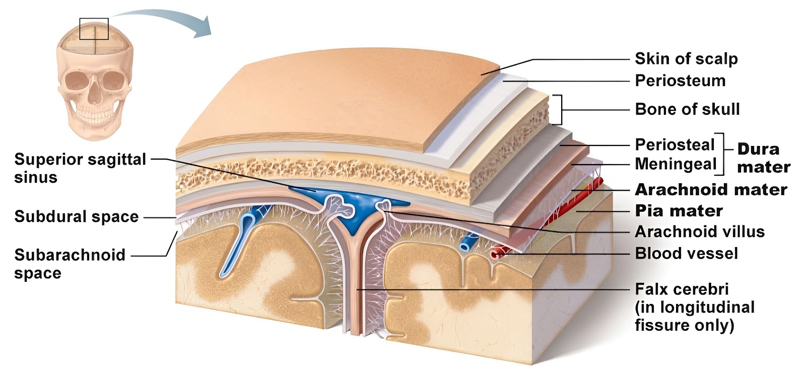
Functions of meninges are; It protects and supports the central nervous system, It acts as a shock absorber, It supplies blood vessels deliver the blood to CNS tissue and its special function is it produces several spinal fluids. The meninges and CSF act as a shock absorber and protect the brain and spinal cord from mechanical injuries. These three sub-layers of meninges are;
Dura mater or membrane : It is the outermost layer of meninges. This layer connects the meninges to the skull and vertebral column. This layer of meninges is composed of tough, white, non-vascular, thick and fibrous connective tissue. This layer of meninges is separated from the underlying arachnoid mater by the subdural space, this space is filled with a serous fluid.
Arachnoid mater or membrane : It is the middle most layer of meninges. This layer of the meninges connects the dura mater and pia mater. This layer of meninges is composed of a non vascular, thin layer of connective tissue having web-like appearance. This layer of meninges is separated from the underlying Pia mater by the narrow subarachnoid space, this space is filled with a cerebrospinal fluid (CSF).
Pia mater or membrane : It is the outermost transparent layer of meninges. This layer of the meninges is in direct contact with and closely covers the cerebral cortex and spinal cord. This layer of meninges is composed of a rich supply of blood vessels, which provide nutrients to nervous tissue. This layer also contains the choroid plexus, a network of capillaries and ependyma that produce cerebrospinal fluid (CSF). This layer of meninges is direct contact with CNS, there is no space.
Brain :
It is the most complicated, highly specialised organ, and important coordinating (command) centre in the body. The human brain is Ectodermal in origin. The average weight of the human brain is is range from 1220 to 1400 grams. The weight of male brain is slightly heavier than the female brain, but it has no relationship with intelligence. The human neural system has about 100 billion neurons, the majority of which occur in the brain.
The brain is the most complex organ in the body and uses 20% of the total oxygen we breathe in. The cerebral blood flow is 750 ml/min, because the brain cannot store Oxygen and nutrients so it requires a constant supply of blood. It is enclosed in a bony case called cranium or brain-box or skull which protects the brain against external injury. The brain is covered by triple layered membranes called meninges. Between the membranes and the brain and also inside the brain, there is a lymphatic fluid, called cerebrospinal fluid (CSF), which also protects the brain.
The study of all aspects of the brain is called Encephalology. Growth of the cranial skeleton is closely related to the growth of the brain. If the brain fails to develop in an embryo, then the cranial cavity is also deformed and this clinical condition is called Anencephaly. The brain is formed of two types of nervous tissue. Grey matter on the outer side and White matter on the inner side. All the parts of the brain work together, but each part has its own special properties. The brain can be divided into three main parts viz;
Fore-brain,
Midbrain, and
Hind-brain.

Fore-brain :
It is also known as Pros-encephalon (from Greek; en meaning in, and cephalon meaning brain). It is the largest part of the brain as compared to the other three parts. It is the uppermost part of the brain.
The Fore-brain include;
Olfactory lobes,
Cerebrum, and
Diencephalon.
Olfactory lobes :
The anterior part of the brain is formed by a pair of short club-shaped structures, the olfactory lobes. It is also known as Rhinencephalon. Each lobe consists of two parts; Anterior olfactory bulb, and Posterior olfactory tract. They are fully covered by the cerebral hemispheres and are, therefore; only visible in the ventral view of the brain. A pair of olfactory nerves arises from the olfactory lobes. Olfactory lobes are concerned with the sense of smell.
Cerebrum :
It is also known as Telencephalon. The cerebrum is the largest, making up about 85% of the total brain. And most complex of all the parts of the human brain. The cerebellum is composed of, Left cerebral hemisphere, and the Right cerebral hemisphere. A very deep fissure, the longitudinal fissure, separates the two cerebral hemispheres. The two hemispheres internally connected to each other by a thick band of myelinated nerve fibres called Corpus callosum or Heart body, it is 10 cm long, and anterior end of Corpus callosum is called Genu, while posterior end of Corpus callosum is called Splenium. The Splenium is continuous with another tract called Fornix.
The outer surface of the cerebrum is called the cerebral cortex while the deep Inner part is the cerebral medulla. The cerebral cortex has an outer thin region composed of grey matter and inner medulla composed of white matter. The surface of the cortex is greatly folded. The upward folds called gyri, alternate with the downward grooves called sulci. Some sulci go deep into the brain, these are called Fissures.
Each cerebral hemisphere of the cerebrum is divided into four lobes by tree Sulcus or fissures, viz; Frontal lobes, Parietal lobes, Temporal lobes, and Occipital lobes. The central sulcus separates the frontal lobe from the parietal lobe. The lateral or sylvian sulcus separates the frontal lobe from the temporal lobe. The parieto-occipital sulcus separates the parietal lobe from the occipital lobe. Since these three sulci are not complete the lobes are not clearly demarcated from each other. A fifth median lobe called insula or insular cortex is folded deep within the lateral sulcus.
Frontal lobes :
Premotor area : It is for involuntary movements.
Motor (or pre-central) area : They are for voluntary movements.
Broca's area : They are for speech, movement of vocal cords.
Association area : They are for coordination of movements, interpretation and storage
Parietal lobes :
Gustatory area : It is for the sense of taste.
Somesthetic (or post-central or Sensory) area : It is for general sensations like pain, touch, temperature etc.
Temporal lobes :
Wernicke's (or intelligence) area : It is for understanding speech.
Olfactory (or smell) area : It is for the sense of smell.
Auditory (or hearing) area : It is for the sense of hearing.
Occipital lobes :
Visual area : It is for the sense of vision.

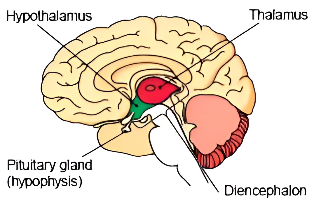
Diencephalon :
It is the part of the forebrain which lies above the midbrain,below the corpus callosum, and between the lower parts of the two cerebral hemispheres. It encloses a single cavity termed third ventricle or Diocoel communicates with the two lateral ventricles of cerebrum through a narrow opening called Foramen of Monro. Diencephalon is the part of the forebrain that contains;
Epi-thalamus :
According to the typical division of the brain, It is the roof of diencephalon of the forebrain. Epithalamus is fused with overlined. pia mater to form anterior choroid plexus (ACP) which produces CSF. On the anterior side, epithalamus has Pineal Gland (Epiphysis) or Third Eye or Biological Clock or "Seat of Soul". It produces two hormones i.e. Melatonin (Level is more during Scotophase or dark phase), and Serotonin (level is more during Photophase or light phase).
Thalamus :
According to the typical division of the brain, It is the walls of the diencephalon of the forebrain. Thalamus is an egg-shaped mass of grey matter. It also contains white matter to some extent. On the superior surface, thalamus is covered by a thin layer of white matter. is called stratum zonale. Thalami are relay centres. These relay sensory impulses except that of smell.
Hypo-thalamus :
According to the typical division of the brain, It is the floor of the diencephalon of the forebrain. The floor of the hypothalamus continues as a downward projection called hypophyseal stalk or infundibulum which connects it to the hypophysis (pituitary gland) both physically and functionally by secretion of neurotransmitters. It is that area of the brain where nervous and endocrine systems are integrated. so it is a neuroendocrine structure. Therefore, it performs both; Nervous functions, as well as Endocrine functions.
Nervous functions : It is highly vascular and regulates behavior essential for survival of species i.e., feeding, fighting, fleeing, mating (sex desire/ libido). It has; Hunger centre (lateral hypothalamus), Satiety centre (ventromedial hypothalamus), Thirst centre, and Osmoregulatory centre. It regulates body temperature i.e., It is a Thermoregulatory centre. It regulates emotional reactions.
Endocrine functions : Hypothalamus has "nuclei which synthesise neurohormones. Hypothalamic neurohormones reach anterior pituitary through Hypothalamo -Hypophyseal Portal System and reach posterior pituitary through axons of hypothalamic neurons. These neurohormones regulate the activity of the pituitary gland. All hormones of hypothalamus are peptide hormones.
Midbrain :
It is also known as Mes-encephalon. It is the smallest part of the brain as compared to the other three parts. It is the middle most part of the brain. located between diencephalon and the pons varolii. The Midbrain include;
Corpora quadrigemina, and
Crura cerebri.
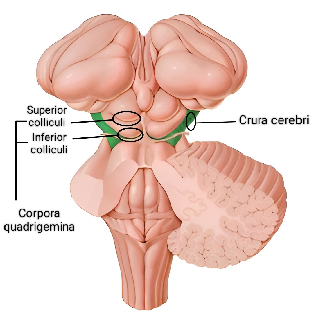
Corpora quadrigemina :
The corpora quadrigemina are four rounded elevations (extremely small swellings) on the dorsal surface of the midbrain. It is located at back of your brain stem (ie. midbrain, at posterior part).
When you automatically look towards a sudden flash of light, or turn towards a startling sound, or when someone calls your name, These reflexive actions are mediated by corpora quadrigemina.
The quadruple (multiple of 4) bodies of corpora quadrigemina is called colliculi. The entire region of corpora quadrigemina, consisting of the four colliculi, is known as the tectum, or tectal plate. There are two superior and two inferior colliculi, with one of each sitting on either side of the midline. The superior colliculi play an important role in the visual pathway, while the inferior colliculi are important in the auditory pathway.
Each quadruple body is related to a bundle of nerve fibres called the brachium. The superior brachium establishes the connection between the two superior colliculi, the lateral geniculate body and the optic tract. The inferior brachium links the two inferior colliculi with the medial geniculate body, which in turn is connected to the auditory cortex. The geniculate bodies are relay centres which play a role in vision and hearing.
Crura cerebri :
It is also known as cerebral peduncle or crus cerebri or crura cerebri. The anterior region of the midbrain is made up of two longitudinal bands of myelinated nerve fibres. They relay impulses back and forth between; Cerebrum, Cerebellum, Pons varolii, and Medulla oblongata.
Near the centre of the midbrain is the mass of grey matter scattered within the white matter; it is called the red nucleus or nucleus rubber. The functions of the red nucleus are; Controlling posture and Muscle tone, Modifying some motor activity, and Motor coordination.
Hind-brain :
It is the second largest part of the brain as compared to the other three parts. It is also known as Rhomb-encephalon. It is the lower most part (posterior region) of the brain. The Hind-brain include;
Cerebellum,
Pons varolii, and
Medulla oblongata.
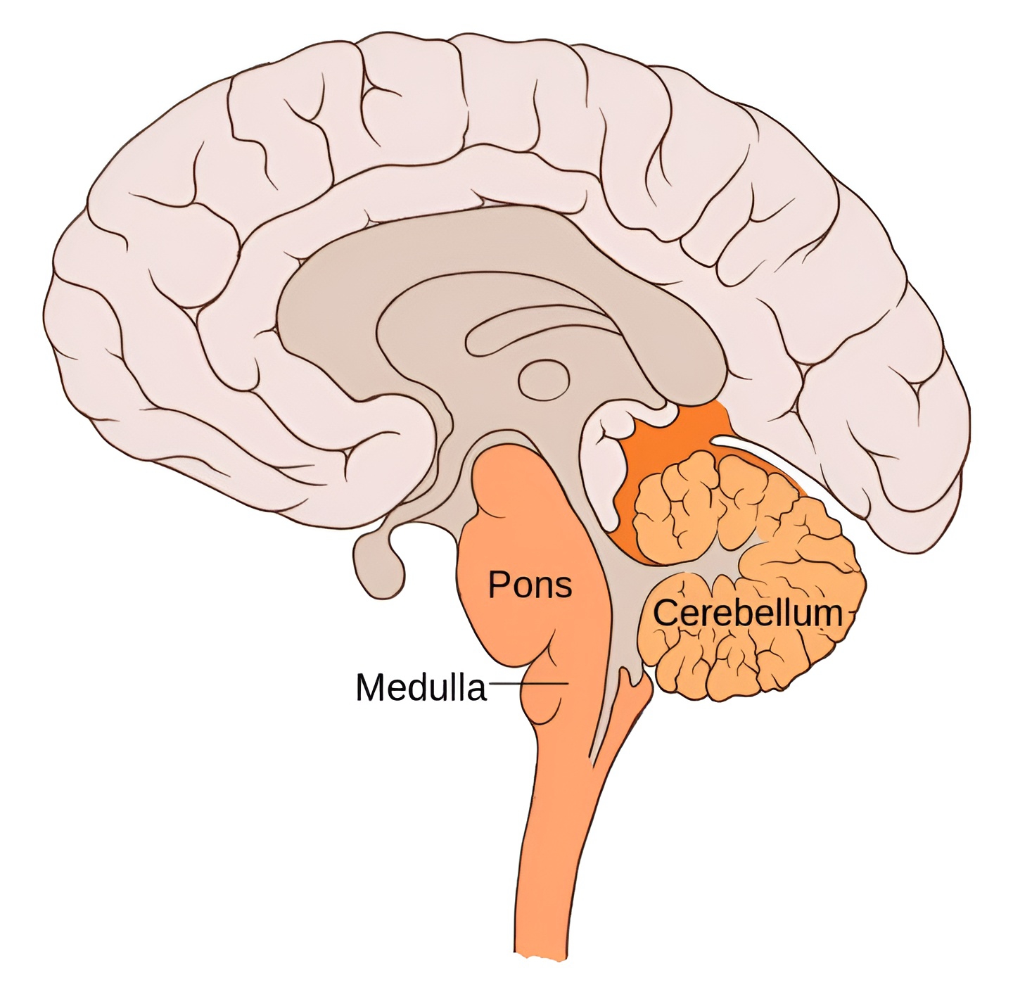
Cerebellum :
It is the second largest part of the human brain (means simply "little cerebrum") or little brain. It is well developed in the human brain. It consists of two lateral cerebellar hemispheres and a central worm shaped part, called the vermis. Like the cerebrum, the cerebellum has its grey matter on the outside, comprising three layers of cells and fibres.
The middle layer contains characteristically large flask shaped Purkinje cells. The Purkinje's cells rank among the most complex of all neurons. The cerebellum also has Golgi cells, basket cells and granule cells. Gyri and sulci of the cerebellum are more symmetrical. A cross section of the cerebellar hemispheres shows a branching tree-like arrangement of grey and white matter called the arbour vitae (or Tree of life).
Some important functions of the cerebrum are;
Controls of rapid muscular activities, such as running, typing and even talking.
Maintenance of balance and equilibrium, Maintenance of muscle tone.
All activities of the cerebellum are involuntary, but may involve learning in their early stages. Alcohol affects the cerebellum.
Pons varolii :
It is situated in front of the cerebellum below the midbrain and above the medulla oblongata. It consists mainly of nerve fibres which form a bridge (pons-bridge) between the two hemispheres of the cerebellum and of fibres which pass between the higher levels of the brain and the spinal cord. The pons varolii contains nuclei associated with four pairs of cranial nerves: Trigeminal (V) nerves, Abducens (VI) nerves, Facial (VII) nerves, and Vestibulocochlear (VIII) nerves. Some important functions of pons varolii are;
Pons varolii relays impulses. between the medulla oblongata and the more superior part of the brain, between the hemispheres of the cerebellum and between the cerebrum and cerebellum.
It has an apneustic area and pneumotaxic area for regulating respiration.
Medulla oblongata :
The medulla oblongata is the posterior conical part of the brain and continues as the spinal cord. It has inner grey matter and outer white matter. The cavity of medulla is called IVth ventricle or metacoel. Its roof is fused with Pia mater, to form posterior choroid plexus for secretion of CSF. Three openings in the roof of the fourth ventricle are; A pair of lateral apertures (foramina of Luschka), and A median aperture (foramen of Magendie). They allow cerebrospinal fluid to move upward to the subarachnoid space that surrounds the brain and spinal cord.
The Medulla oblongata contains nuclei associated with five pairs of cranial nerves; Vestibulocochlear (VIII) nerves, Glossopharyngeal (IX) nerves, Vagus (X) nerves, Accessory (XI) nerves, and Hypoglossal (XII) nerves.
Some functions of medulla oblongata are;
It controls involuntary vital functions centres like; Heart beat, Respiration, Vasomotor activities, and Peristalsis, etc.
It also controls non-vital reflex activities centres like; Coughing, Sneezing, Swallowing, and Vomiting, etc.
Spinal Cord :
It is also known as Myelon. It is part of the central nervous system (CNS). It forms the lower extension of the medulla oblongata of the brain. It is present inside the neural canal or spinal canal of the vertebral column. Externally, the spinal cord appears as long cylindrical rod of 42 to 45 cm long and 2.0 to 2.5 cm broad.

During embryonic life, the spinal cord and vertebral column are of equal length but after birth, the vertebral column grows at a faster rate. Spinal cord begins at the 1st cervical vertebra and terminates to a point called Conus medullaris at the level of L1 to L2 vertebrae. Conus medullaris is connected to the coccygeal and vertebral column by a fibrous connective tissue derived from pia mater called Filum terminale.
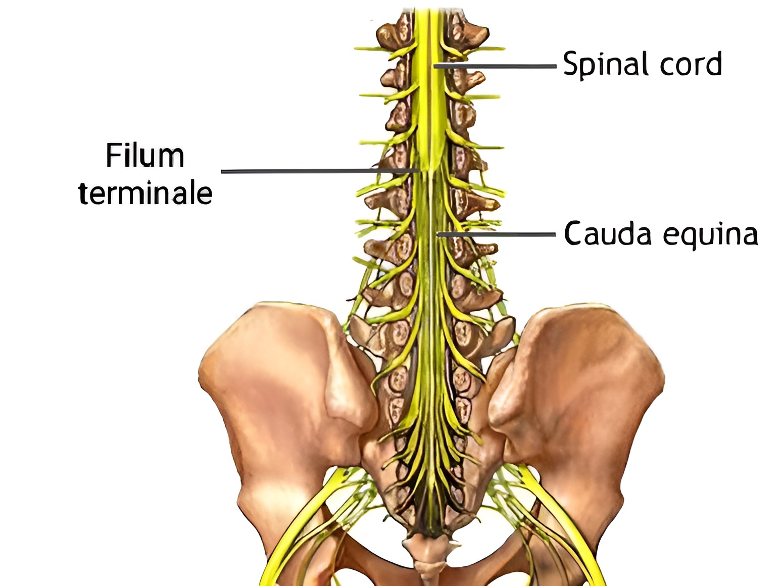
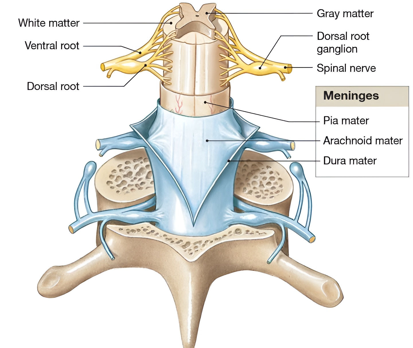
Like the brain, it is covered and protected by bony covering (skeleton) and membranes called meninges (dura mater, arachnoid mater and pia mater). The Cerebrospinal Fluid (CSF) secreted by pia mater, forms a fluid cushion around the spinal cord and within it inside the central canal. Spinal cord shows two swellings along its length called; Cervical swelling, and Lumbar swelling.
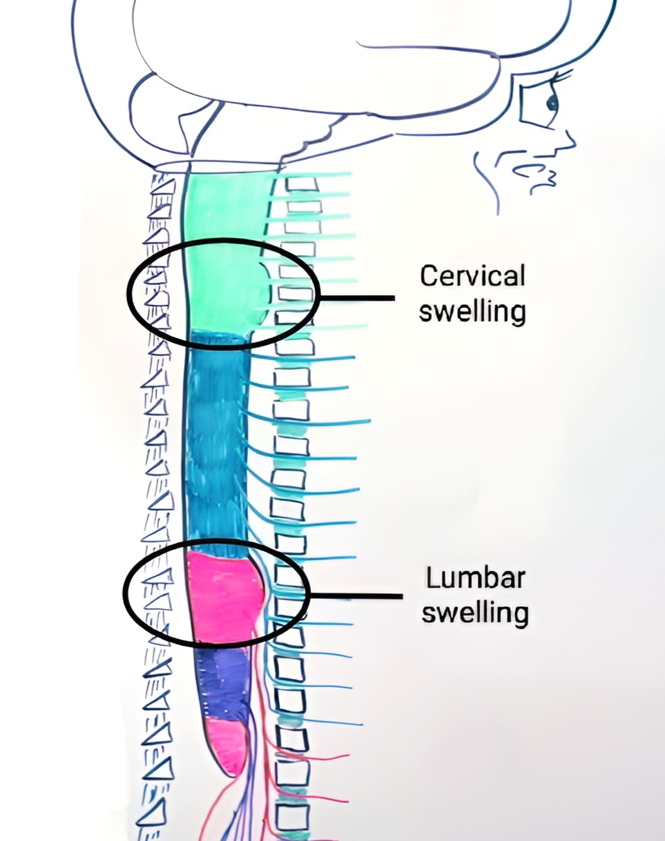
31 pairs of spinal nerves are present in humans, these nerves arise from lateral sides of the spinal cord. These nerves are concentrated in the region of cervical and lumbar swelling and around the conus medullaris. The bunch of nerves in the hind part of the spinal cord, along with the filum terminale, appear like a horse's tail, so called Cauda equina.
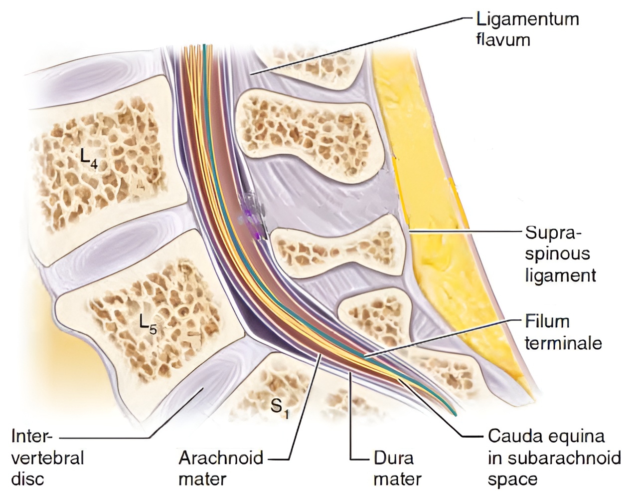
Spinal segment refers to a region of spinal cord from where one pair of spinal nerves develops. Each spinal nerve is attached to the spinal cord by two roots, viz; Posterior dorsal sensory root (it is connected to root ganglion), and Anterior ventral motor root. Anterior and posterior roots join to form spinal nerves, which come out of the vertebral column through intervertebral foramen. After coming out from intervertebral foramen, each spinal nerve divides into three branches called Rami,viz; Dorsal Ramus, Ventral Ramus, and Recurrent (or Meningeal) Ramus.
T.S. of spinal cord :
The spinal cord is dorsoventrally flattened due to the presence of deep, narrow posterior median fissure and shallow, broad anterior median fissure. The fissures divide the spinal cord incompletely into a right and left side, A central canal can be seen in the centre.

It has both grey matter as well as white matter, Grey matter is inside and white matter is outside. Grey matter is arranged in the form of an H or butterfly shaped. The fissures divide the grey matter into six horns, namely, two Dorsal horns, two Lateral horns, and two Ventral horns. While the white matter is divisible into six columns or funiculi, namely, two Dorsal funiculi, two Lateral funiculi, and two Ventral funiculi. The dorsal and ventral horns extend out of the spinal cord as dorsal root and ventral root of the spinal cord respectively. Of these, the dorsal root is connected to the dorsal root ganglion.

The lateral horns have neurons of the autonomic nervous system (ANS). The white matter consists mainly of bundles of myelinated nerve fibre called ascending and descending tracts. The ascending tracts conduct sensory impulses from the spinal cord to the brain and these lie in the dorsal column/funiculi, While the descending tracts conduct motor impulses from brain to the lateral and ventral funiculi of spinal cord.
The spinal cord is the main centre for the most reflex actions. It provides a pathway for conduction of sensory and motor impulses to and from the brain. It provides a nervous connection to many parts of the body.
Peripheral Nervous System (PNS) :
The peripheral nervous system (PNS) connects the central nervous system (CNS) to the different parts of the body having receptors and effectors. The PNS is divided into two divisions called Somatic Neural System (SNS) and Autonomic Neural System (ANS).
Based on their function, the peripheral nervous are divided into three types; Afferent or sensory neuron (Transmit messages towards the CNS), Efferent or motor neuron (Transmit messages away from the CNS), and Interneurons or Association neuron (perform both function). Based on their connection to the CNS, the peripheral nerves are classified into two main types; Cranial and Spinal nerves. Both cranial and spinal nerves can have sensory, motor, or mixed functions.
Somatic Neural System (SNS) :
The somatic neural system relays impulses from the CNS to skeletal muscles. Therefore, it is also known as the Voluntary nervous system.
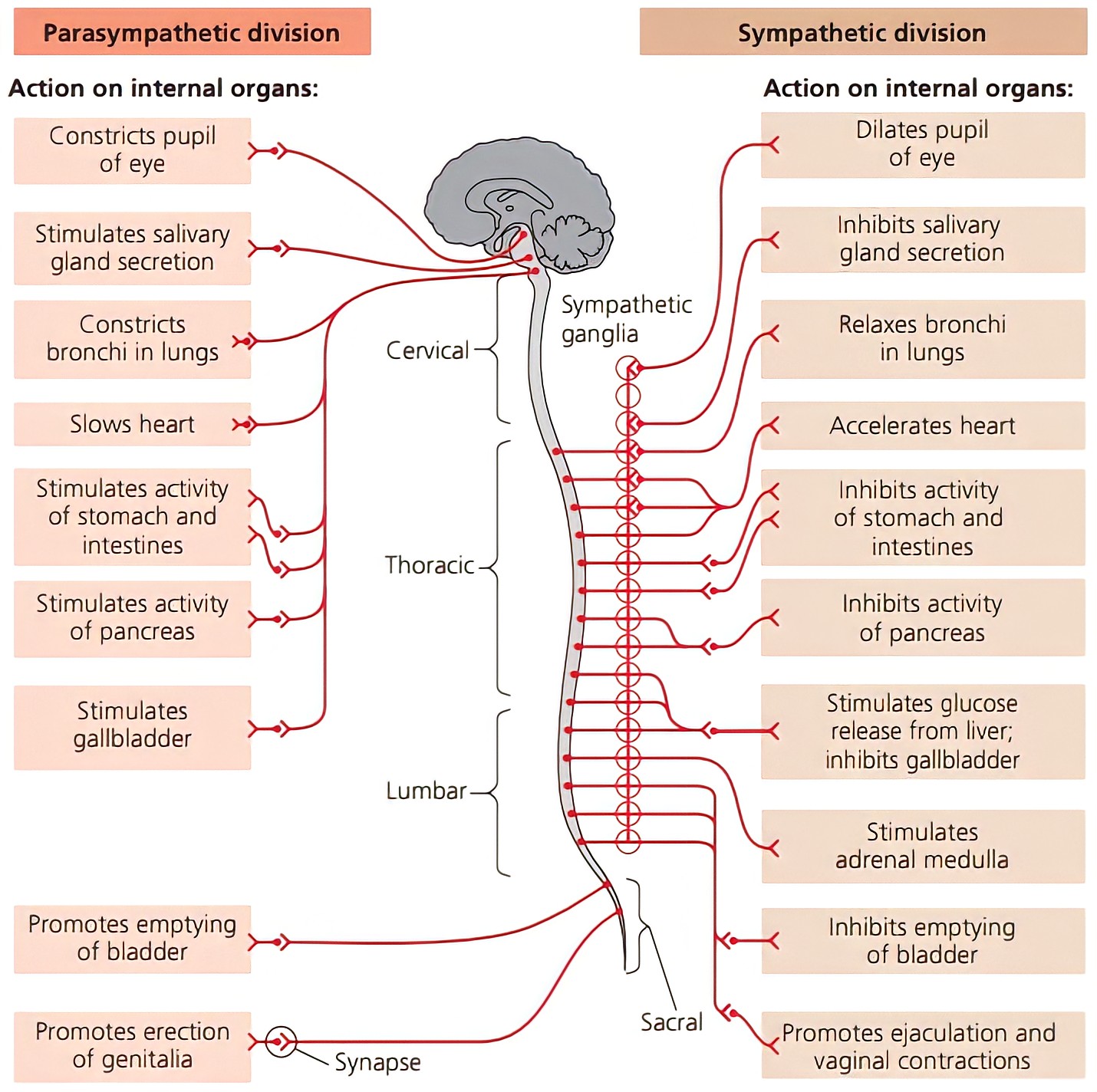
Autonomic Neural System (ANS) :
Autonomic nervous system transmits impulses from CNS to the involuntary organs and smooth muscles of the body. Therefore, it is also known as the Visceral or Involuntary or Vegetative nervous system. ANS consists of a special set of peripheral nerves that regulate the activities of involuntary organs like; Hearing a sudden loud noise, Cardiac muscles, Smooth muscles, and Glands, etc. In this, impulses are conducted from the CNS like this;
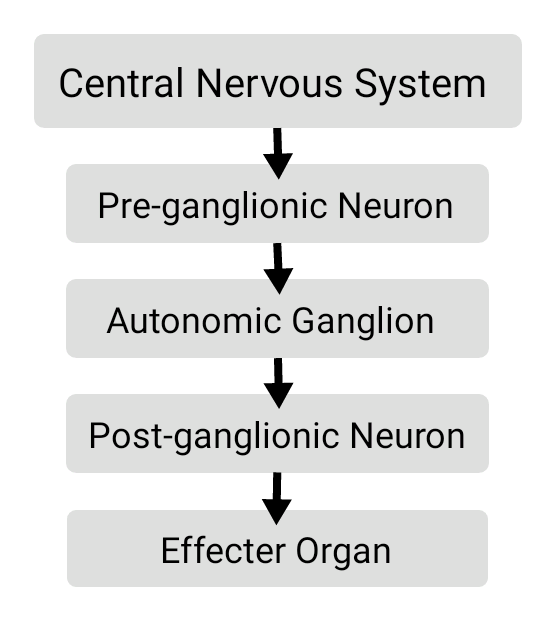
Autonomic nervous system is subdivided into;
Sympathetic Nervous System,
Parasympathetic Nervous System, and
Enteric Nervous System.
Sympathetic and Parasympathetic nervous systems are antagonistic to each other.
Enteric Nervous System :
The enteric nervous system or intrinsic nervous system is a mesh-like system of neurons that governs the function of the gastrointestinal tract.
Cranial nerves :
These nerves are connected to the brain of the central nervous system (i.e. originate from the brain). 12 pairs of cranial nerves are present in Amniotes (Reptiles / Birds / Mammals), while 10 pairs of cranial nerves are present in Anamniotes (Fishes / Amphibians). Roman numbering I to XII is used to denote them. The pairs of cranial nerve are;
Olfactory nerves,
Optic nerves,
Oculomotor nerves,
Trochlear nerves,
Trigeminal nerves,
Abducens nerves,
Facial nerves,
Auditory nerves,
Glossopharyngeal nerves,
Vagus nerves,
Accessory nerves, and
Hypoglossal nerves.
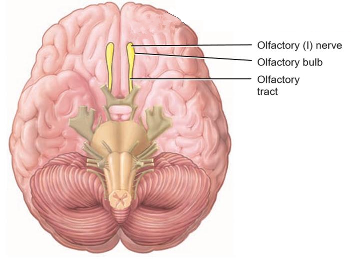
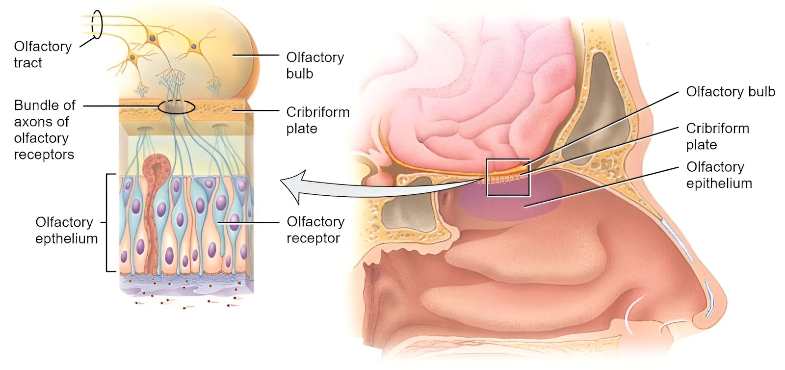
Olfactory nerves :
It is denoted by the Roman number I. It is entirely a sensory cranial nerve. It originated from an olfactory bulb. It is innervated with epithelium of the nose, the olfactory epithelium occupies the superior part of the nasal cavity. This is responsible for the sense of smell.

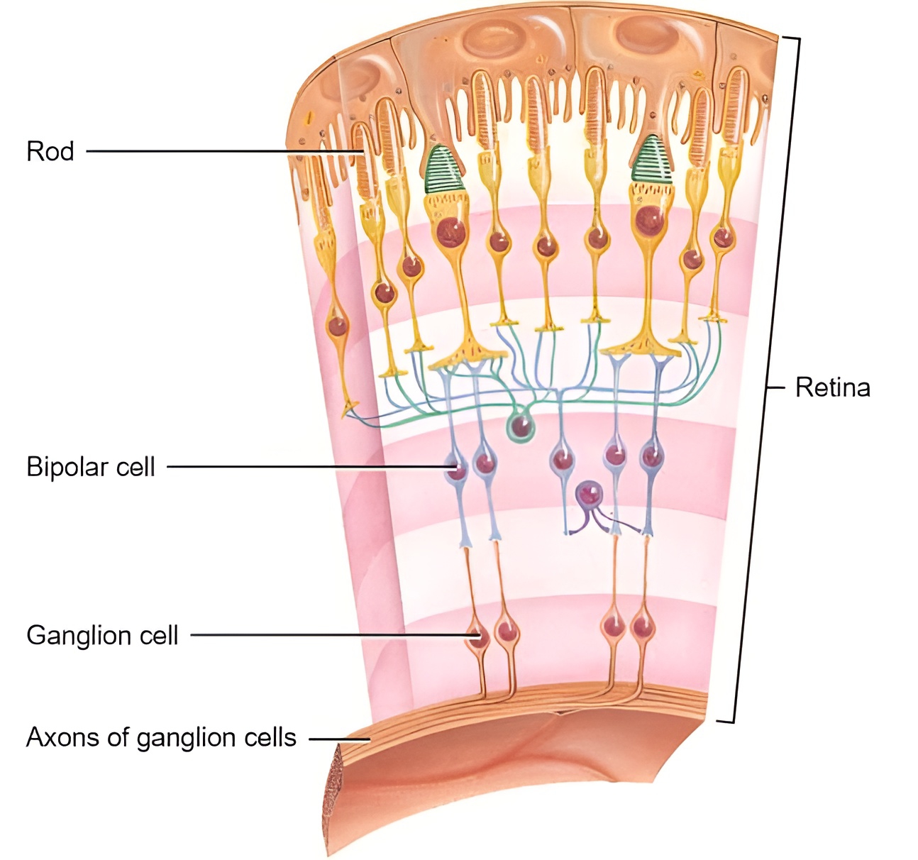
Optic nerves :
It is denoted by the Roman number II. It is entirely a sensory cranial nerve. It originated from an diencephalon. It is innervated with the retina of eyes, the retina of each eye joins to form an optic nerve, which passes through the optic foramen. This is responsible for a sense of vision.

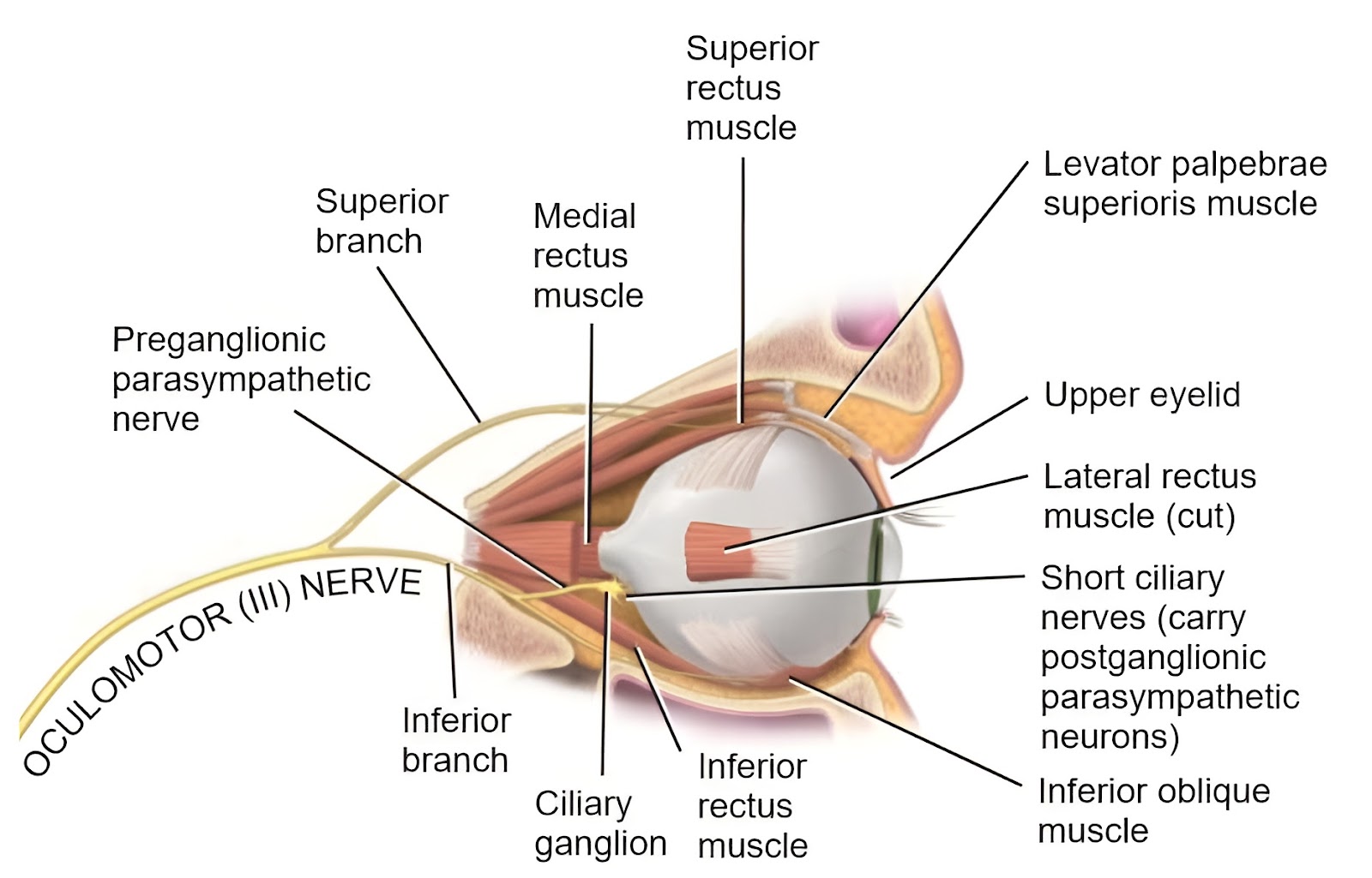
Oculomotor nerves :
It is denoted by the Roman number III. It is entirely motor cranial nerve. It originated from the floor of midbrain. It is innervated with Eye muscles (4 of 6 eye muscles). These somatic motor neurons control movements of the eyeball and upper eyelid. This is responsible for movement of the eyeball.
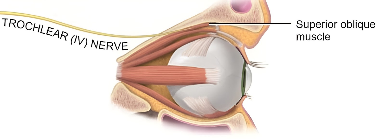
Trochlear nerves :
It is denoted by the Roman number IV. And it is also called Pathetic nerves. It is entirely motor cranial nerve. It originated from the floor of midbrain. It is innervated with Eye muscles (1 of 6 eye muscles, forehead scalp), it is the smallest and thinnest of all the cranial nerves. This is responsible for movement of the eyeball.
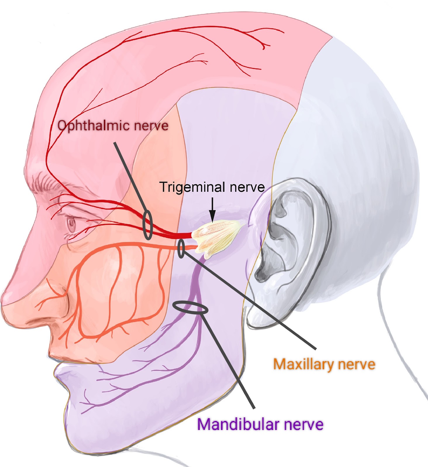
Trigeminal nerves :
It is denoted by the Roman number V. And it is also known as Dentist's nerves. It is an entirely mixed cranial nerve. It originated from the ventral side of pons varolii. It is the largest and thickest of all the cranial nerves. Trigeminal nerve has three branches;
Ophthalmic nerve : It is entirely a sensory cranial nerve. It is innervated with; Nasal cavity, Upper eyelids, Forehead, Scalp, Conjunctiva, and Lacrimal gland.
Maxillary nerve : It is entirely a sensory cranial nerve. It is innervated with; Mucosa of nose, Palate, Upper teeth, Upper lip, Lower eye, and Lid parts of pharynx.
Mandibular nerve : It is an entirely mixed cranial nerve. It is innervated with; Lower teeth, Skin over mandible cheek, Side of head in front ear, and Muscles of mastication.
This is responsible for the sensation of skin touch, taste, jaw moment.
Abducens nerves :
It is denoted by the Roman number VI. It is entirely motor cranial nerve. It originated from pons varolii. It is innervated with muscles of the eyeball, internal rectus muscle. This is responsible for movement of the eyeball.

Facial nerves :
It is denoted by the Roman number VII. It is an entirely mixed cranial nerve. It is originated from pons varolii It is innervated with; Facial, Scalp and neck muscles, Lacrimal, Sublingual, Submandibula, Nasal, and Palatine glands. This is responsible for; Facial expression, Movement of neck, Secretion of tears, Taste, and Salivary secretion.

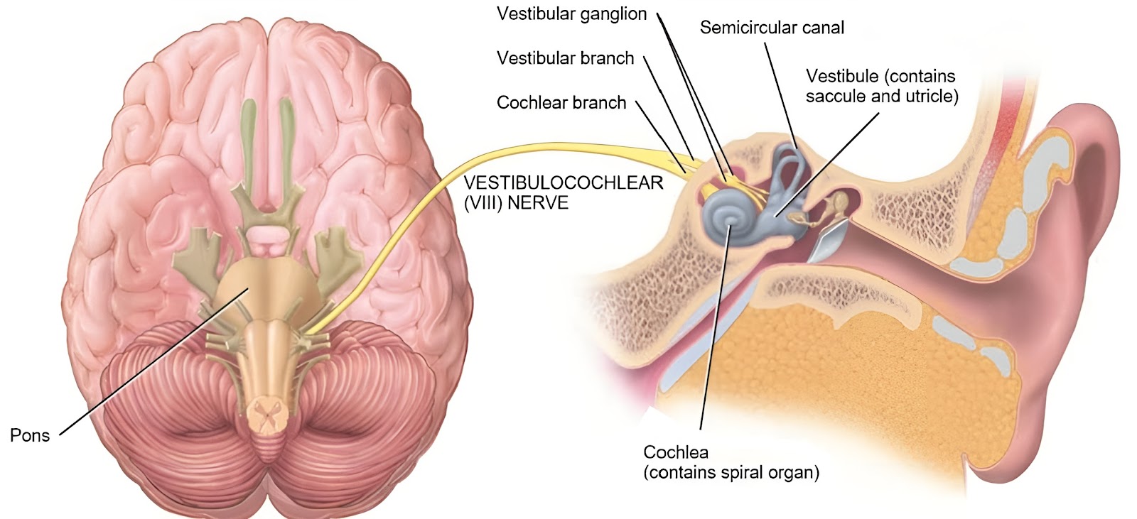
Auditory nerves :
It is denoted by the Roman number VIII. It is also known as Vestibulocochlear Nerve or Acoustic nerve. It is entirely a sensory cranial nerve. It originated from pons varolii. Auditory nerve has two branches, the Vestibular branch, and the Cochlear branch. It is innervated with the internal ear. This is responsible for hearing (Cochlear branch) and equilibrium (Vestibular branch).
Glossopharyngeal nerves :
It is denoted by the Roman number IX. It is an entirely mixed cranial nerve. It originated from the side of medulla oblongata It is innervated with; Pharynx, Tongue, and Salivary glands. This is responsible for; Taste, Salivation, and Swallowing.

Vagus nerves :
It is denoted by the Roman number X. It is an entirely mixed cranial nerve. It originated from the side of medulla oblongata. It is the longest of all the cranial nerves, It is the only cranial nerve which crosses the neck region, and it has a maximum number of branches which is widely distributed in the body. It is innervated with; Larynx, Trachea, Pharynx, Alimentary canal, Heart, Lungs, Pancreas, and Blood vessels. This is responsible for Visceral sensations and visceral movements like; Breathing cardiac, Slowing, Gastric and Pancreatic secretion, and Gastrointestinal movements.

Accessory nerves :
It is denoted by the Roman number XI. It is entirely motor cranial nerve. It originated from the side of medulla oblongata. Historically it has been divided into two parts; Cranial accessory nerve, and Spinal accessory nerve. It is innervated with; Larynx, Pharynx, Neck, and Shoulder. This is responsible for movements of; Larynx, Pharynx, Neck, and Shoulder.

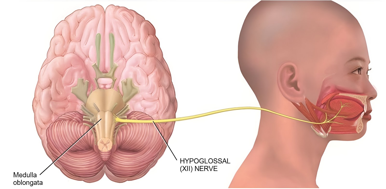
Hypoglossal nerves :
It is denoted by the Roman number XII. It is entirely a motor cranial nerve. It originated from the side of medulla oblongata. It is innervated with tongue muscles. This is responsible for movement of the tongue.
Spinal nerves :
These nerves are connected to the spinal cord of the central nervous system (i.e. originate from the spinal cord). 31 pairs of spinal nerves are present in humans, these nerves arise from lateral sides of the spinal cord. All spinal nerves are of the mixed type (ie. they have some nerve fibre as sensory and motor).
These nerves are concentrated in the region of cervical and lumbar swelling and around the conus medullaris. The bunch of nerves in the hind part of the spinal cord, along with the filum terminale, appear like a horse's tail, so called Cauda equina. Spinal segment refers to a region of spinal cord from where one pair of spinal nerves develops.
Each spinal nerve is attached to the spinal cord by two roots, viz, Posterior dorsal sensory root (it is connected to root ganglion), and Anterior ventral motor root. Anterior and posterior roots join to form spinal nerves, which come out of the vertebral column through intervertebral foramen. After coming out from intervertebral foramen, each spinal nerve divides into three branches called Rami,viz; Dorsal Ramus, Ventral Ramus, and Recurrent (or Meningeal) Ramus. Number and types of spinal nerves are;
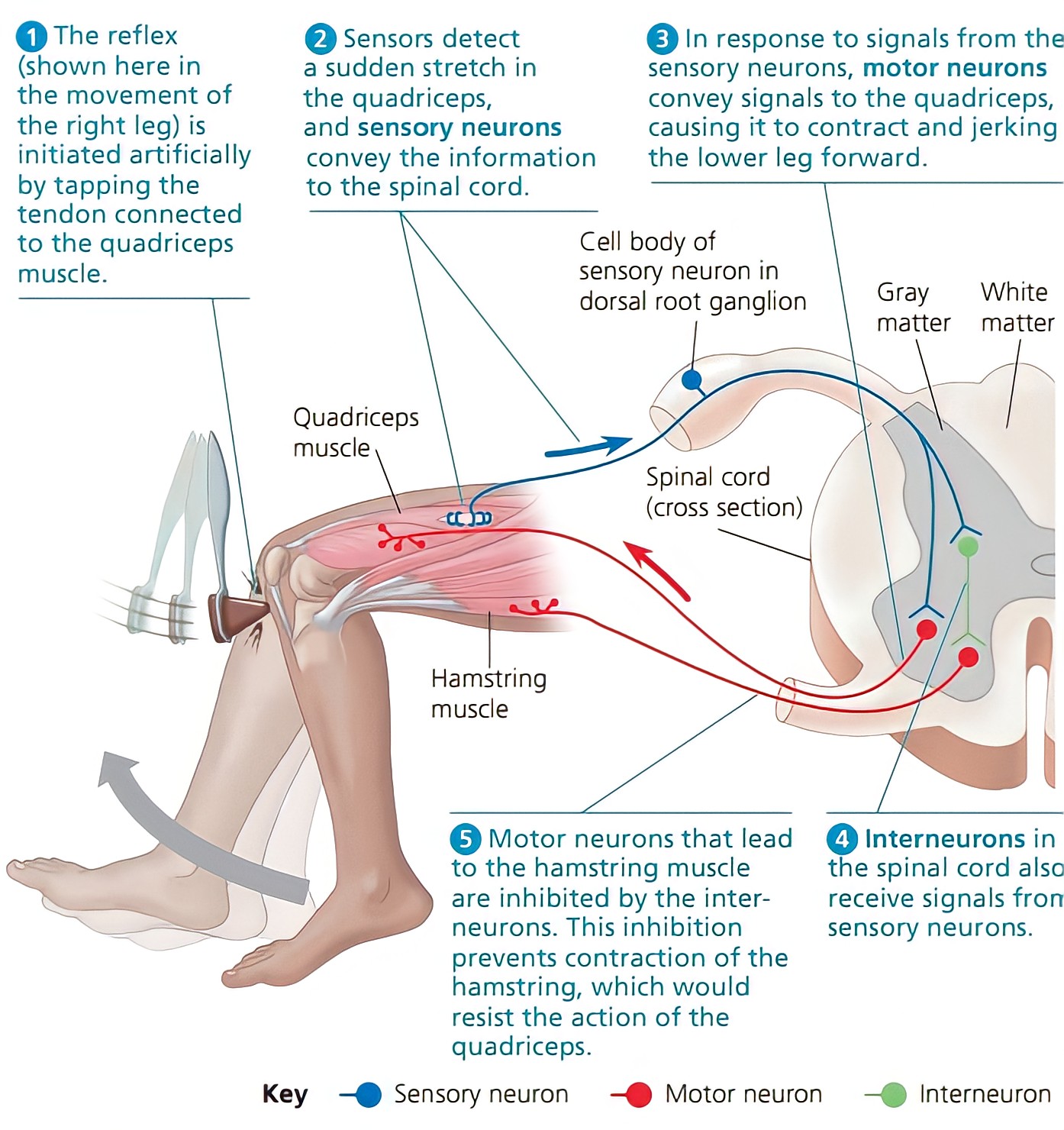
Reflex Action :
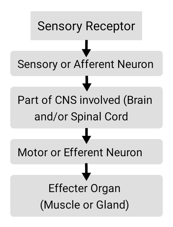
It is a sudden, spontaneous, automatic, involuntary response to a stimulus acting on the receptor. The response to stimulus is said to be involuntary as it is carried out without any conscious effort by the brain. The reflex action was first observed by Marshall Hall in the year 1833. During reflex action the impulse travels through a path known as the Reflex arc. The reflex arc typically consists of five components, and impulse travel through reflex arc as;
Based on previous experiences, reflex actions are of two types; Unconditional or Inborn or Inherited reflexes, and Conditional or Acquired reflexes. Based on the part of CNS involved in reflex arc, reflex actions are of two types; Cranial reflexes, and Spinal reflexes. Based on the number of synopsis involved in reflex arc, reflex actions are of two types; Simple Mono-sympathetic reflexes, and Complex Poly-sympathetic reflexes.
Some important examples of reflex action are; Narrowing of the pupil of eye on seeing bright light, Withdrawal of limbs when it touches hot/cold object, Quick closing of eyelids when a flying object suddenly approaches the eye, Coughing, Sneezing, Yawning, Shivering, Opening of the mouth on hearing a sudden loud noise, and Knee jerk reflex (shown below).
Brain stem :
The midbrain, pons varolii and medulla oblongata are collectively called the brain stem, connecting the forebrain and spinal cord.

Ventricles of the Brain :
The ventricles are the fluid filled spaces inside the brain, simply brain cavities. Ependymal cells line the brain cavities. The ventricular system is composed of; Two lateral ventricles, Third ventricle, and Fourth ventricle.
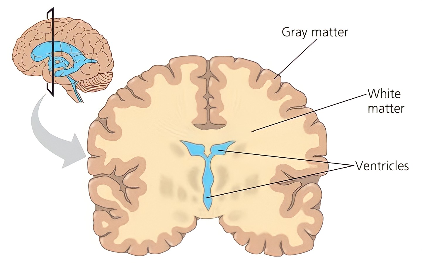
A lateral ventricle lies inside each hemisphere of the cerebrum. Each lateral ventricle is connected to the third ventricle by an interventricular foramen (foramen of Monro). The third ventricle is connected with the fourth ventricle by a narrow channel called Cerebral aqueduct or Aqueduct of Sylvius. The fourth ventricle is continuous with the central canal of the spinal cord.

Three openings in the roof of the fourth ventricle are; A pair of lateral apertures (foramina of Luschka), and A median aperture (foramen of Magendie). They allow cerebrospinal fluid to move upward to the subarachnoid space that surrounds the brain and spinal cord.
Cerebrospinal Fluid (CSF) :
It is lymph-like extracellular, clear colourless slightly alkaline (7.3 pH) fluid with specific gravity 1.005. The volume of cerebrospinal fluid is 80 to 150 ml.
It is continuously produced and reabsorbed. It is continuously secreted by; ACP (Anterior choroid plexus) and PCP (Posterior choroid plexus). The composition of CSF is; Proteins, Amino acids, Potassium ions, Oxygen, Na+, and HCO3, etc. The cerebrospinal fluid is present in; Ventricles of the brain, Central canal of the spinal cord, and Subarachnoid space.
Functions of cerebrospinal fluid are; It provides Oxygen and nutrients to the brain tissue, It collects the metabolic waste material from the brain tissue, It protect brain from desiccation (or it keep brain moist), It acts as a shock absorber, It maintains constant intracranial pressure, and It transport neurotransmitters within brain.
Summary :

Disorders of nervous system :
Alzheimer's disease :
A progressive disease that eventually destroys memory and other important mental functions. Brain cell connections and the cells themselves degenerate and die. It is due to loss of cholinergic and other neurons in the CNS, accumulation of amyloid proteins. There is no cure for Alzheimer's, but treatment slows down the progression of the disease and may improve the quality of life.
Join the conversation