Anatomy and physiology of human ear
Anatomy of human ear :
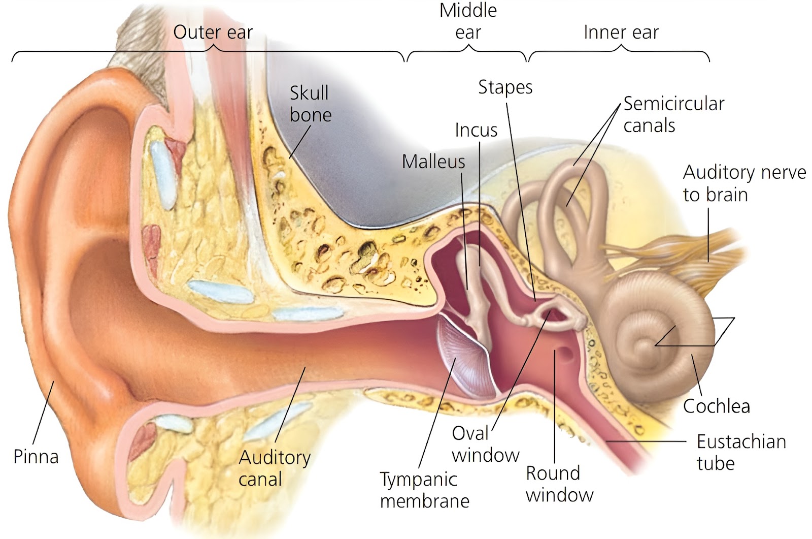
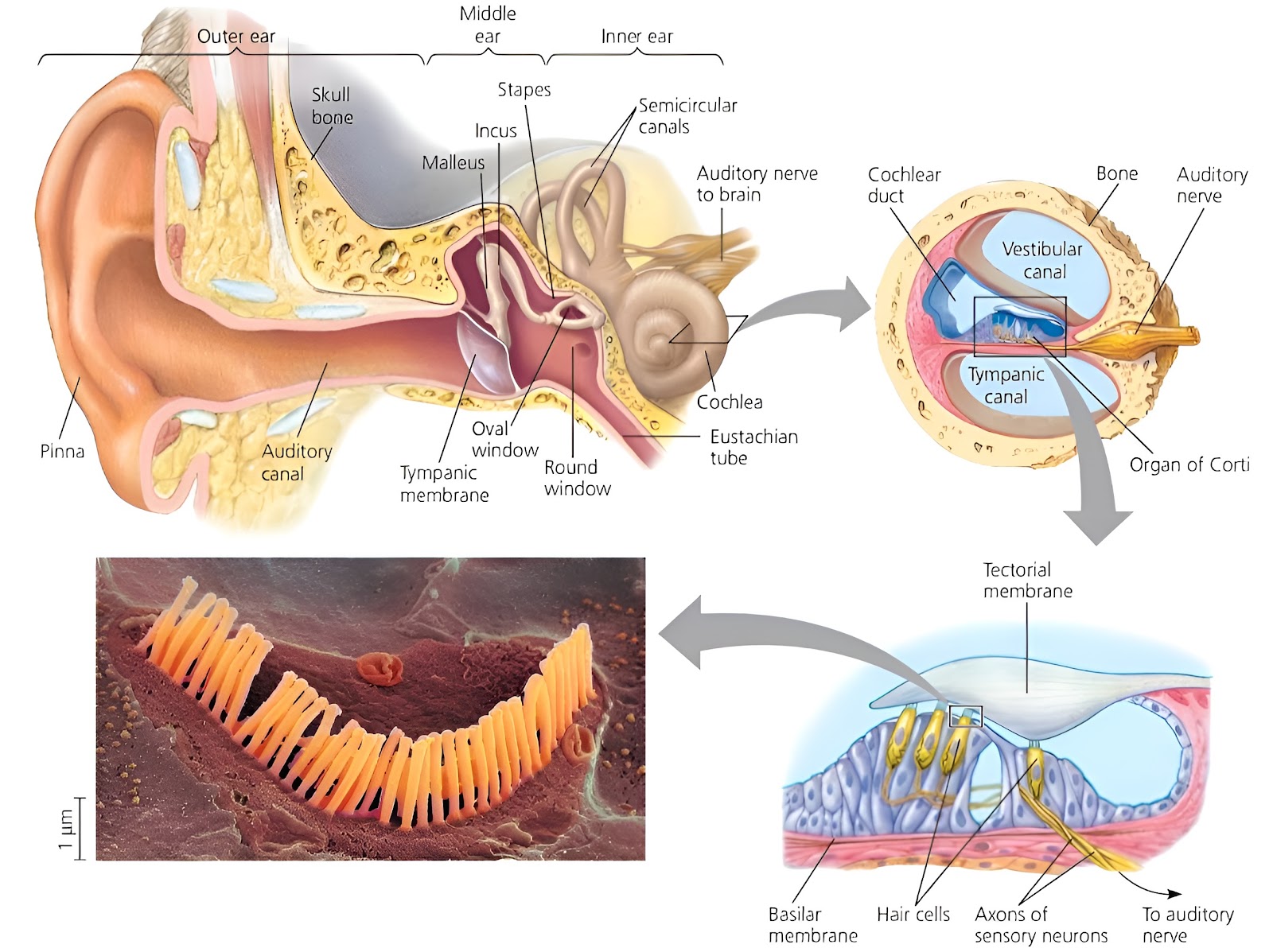
The human ear is called a statoacoustic organ, because it has two main functions; Hearing, and Body balance and equilibrium. Balance and equilibrium is the primary function of the human ear. Anatomically the ear is made up of three parts; External ear, Middle ear, and Internal ear.

External ear :
It is also known as the outer ear. It consists of; Ear pinna (it has auricles), External auditory canal, and Eardrum.
Ear pinna :
It has auricles. Auricle is a flap of yellow elastic cartilage covered by skin. Ligaments and muscles attach the auricle to the head. Its most prominent outer ridge is called the helix. The lobule is the soft pliable part at its lower end composed of fibrous and adipose tissue richly supplied with blood capillaries. It's a characteristic of mammals, except whales, dolphins, and platypus, etc. Many mammals can move their ear pinna in the direction of sound, but in humans it is relatively immobile because the auricular muscles of pinna, which are vestigial. It is sensitive as well as effective in collecting sound waves, and transmits them towards the tympanic membrane. It's difficult to localise sound with one ear.
External auditory canal :
It is also known as External auditory meatus. It is a curved tube about 2.5 cm long. It lies in the temporal bone and leads to the eardrum. It has yellow elastic cartilage. This canal is internally lined by hairy skin (stratified epithelium) and ceruminous or cerumen or wax glands. These are modified sweat glands which secrete a waxy substance, ear wax or cerumen. Ear wax traps foreign particles (i.e. dust, pathogen). It also prevents damage to the delicate skin of the external ear canal by water and insects. Cerumen usually dries up and falls out of the ear canal. Some people produce a large amount of cerumen, which can become impacted and can muffle incoming sounds.
Eardrum :
It is also known as Tympanum or Tympanic membrane. Tympanic membrane is a thin, semi-transparent, almost oval. It is a partition between the external auditory canal and middle ear. It is comprised of all three germ layers (i.e. Ectoderm, Mesoderm, and Endoderm). It is covered by epidermis and lined by simple cuboidal epithelium. The central part of the tympanic membrane is called the umbo. The handle of the malleus is firmly attached to the membrane's internal surface. It vibrates in response to sound waves. For tympanum to vibrate freely in air pressure must be equal on both sides.
Middle ear :
It is also known as Tympanic cavity. The tympanic cavity, filled with air, is connected with the nasopharynx through the Eustachian tube (auditory tube), which is responsible for equalising the pressure on either side of the tympanic membrane. There is a small flexible chain of three small bones called ear or auditory ossicles, these are;
Malleus :
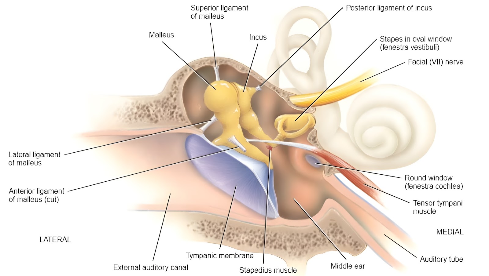
This auditory ossicle is hammer in shape. Malleus is the largest ossicle. The malleus is attached to the tympanic membrane on one side and to the incus on the other side.
Incus :
This auditory ossicle is anvil in shape. The Incus is attached to the malleus on one side and to the stapes on the other side.
Stapes :
This auditory ossicle is stirrup in shape. Stapes is the smallest ossicle. Stapes is also the smallest bone in the body. The stapes are attached to the incus on one side and to the oval membrane on the other side. Covering the fenestra ovalis (oval window) of the inner ear.
Two skeletal muscles are present in a middle ear;
Tensor tympani : It is attached with malleus.
Stapedius : It is attached with stapes. And also it is the smallest muscle in the body.
The middle ear is connected with the inner ear through two small openings closed by the membranes, these openings are;
Fenestra ovalis : It is also known as an oval window. It is covered by the footplate of stapes.
Fenestra rotunda : It is also known as a round window. It is covered by the flexible secondary tympanic membrane.
Internal ear :
It is also known as labyrinth, because of its complicated series of canals. Structurally, it consists of two main divisions; outer Bony labyrinth, and inner Membranous labyrinth.
The Bony labyrinth is a series of cavities in the petrous portion of the temporal bone divided into three areas;
Cochlea,
Vestibule, and
Semicircular canals.
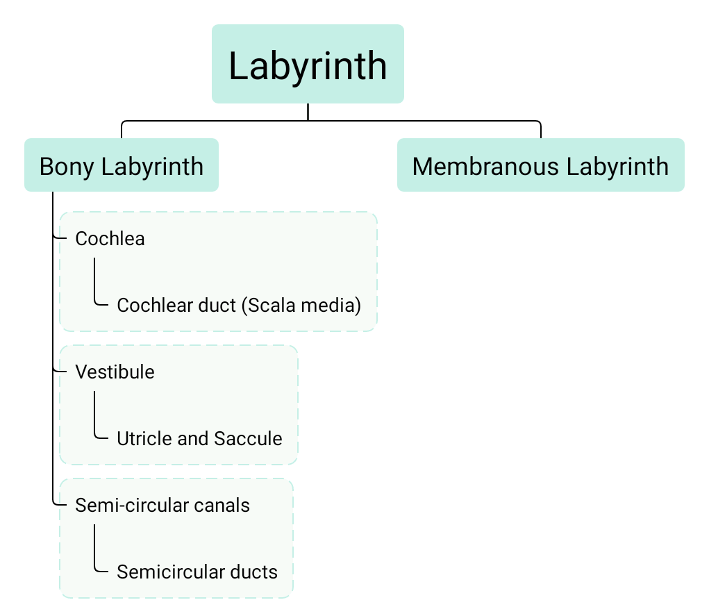
Both vestibule and semicircular canal are together called the vestibular apparatus. They contain receptors for equilibrium. Bony labyrinth is lined with periosteum and contains perilymph.
Cochlea :
Cochlea is a bony spiral canal that resembles a snail's shell in appearance. It makes almost three turns around a central bony core called modiolus. It has a central cochlear duct called scala media. Scala media divides cochlea into two compartments, viz, upper scala vestibuli, and lower scala tympani. Scala vestibuli and scala tympani meet at the tip of the cochlea called helicotrema. Both scala vestibuli and scala tympani are filled with fluid called perilymph, While scala media is filled with fluid called endolymph.
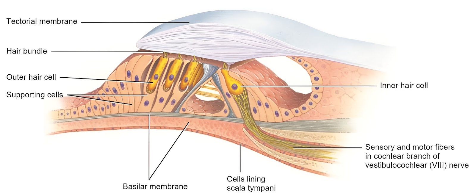
The scala media is the most important canal or channel of the cochlea, it contains receptors for hearing. The base/floor of scala media is formed by basilar membrane and roof of scala media is formed by reissner's membrane. At the basilar membrane of the scala media the actual sense organ for the hearing is present, called the organ of corti.
Vestibule :
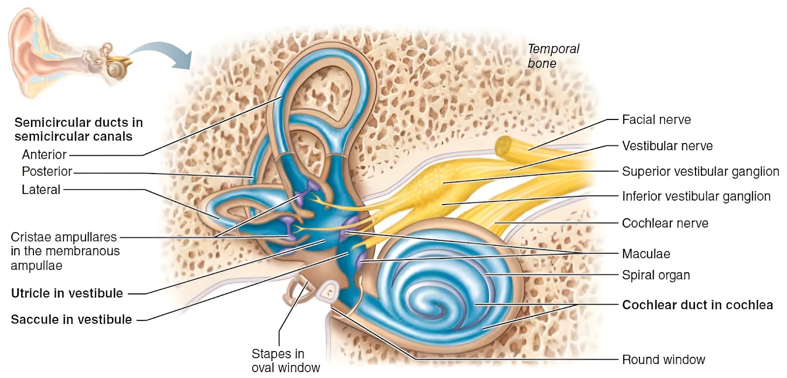
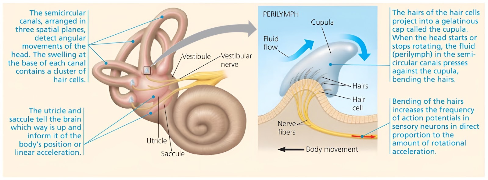
Vestibule is the oval central portion of the bony labyrinth. Membranous labyrinth in the vestibule consists of two sacs called the utricle and the saccule, which are connected by a small duct. The utricle is a dorsally placed structure to which all the three semicircular ducts are connected. The saccule is a ventrally situated structure which is joined with the utricle by a narrow utriculosaccular duct. From this duct a long tube, the ductus endolymphaticus arises. Both the utricle and saccule contain sensory patches called the maculae. A macula comprises sensory and supporting cells similar to those of the crista.
Semicircular canals :
There are three semicircular canals which are mutually perpendicular to each other. Based on their position, they are named as; Anterior semicircular canal, Posterior semicircular canal, and Lateral semicircular canal. These canals are filled with fluid called endolymph. At one end of each canal is a swollen enlargement called the ampulla.
The portions of the membranous labyrinth that lie inside the bony semicircular canals are called the semicircular ducts. Each ampulla contains a sensory patch of cells called the crista. Each crista consists of two kinds of cells, the sensory and supporting cells. The cristae helps in maintaining the body equilibrium. The semicircular ducts arise from the utricle.
Organ of corti :
It is also known as a spiral organ. It is an actual sense organ for hearing. It is present inside scala media on the basilar membrane throughout its length. It has the following types of cells; Sensory hair cells, Supporting cells or Cells of hensen, and Basal cells or Deiter cells. There are two groups of hair cells; The inner hair cells are arranged in a single row, whereas the outer hair cells are arranged in three rows. At the apical tip of each hair cell there are 40 to 80 stereocilia that extend into the endolymph of the cochlear duct. Stereocilia are actually long, hairlike microvilli arranged in several rows of graded height. The Tectorial membrane, a flexible gelatinous membrane, covers the hair cells of the spiral organ. Its properties are to determine the patterns of vibration of sound waves.
Physiology of balance and equilibrium :
The human ear is called a statoacoustic organ, because it has two main functions; Hearing, and Body balance and equilibrium. Balance and equilibrium is the primary function of the human ear. The vestibular apparatus(i.e. semicircular canals and vestibule) is the part of the inner air that is directly involved in helping the body to maintain the balance and equilibrium. The balance and equilibrium of the body is divided into two types on the basis of which part of the vestibular apparatus of the inner ear is involved, viz.
Dynamic equilibrium :
In dynamic equilibrium the semicircular canal of vestibular apparatus is involved, which responds to rotational movement (angular acceleration). Three semicircular canals, each one oriented in a different plane. There is a small chamber at one end of each canal containing hair cells. Whenever the head is moved, the fluid within the canals lags in its motion so that there is relative motion between the walls and the endolymph. This stimulates the hair cells to send impulses back to the brain.
Static equilibrium :
In static equilibrium the utricle and saccule within the vestibule is involved which respond to gravity (linear acceleration). Sacculus and utriculus are interconnected chambers, which are filled with fluid endolymph. On their inner surface there are patches of hair cells to which thousands of tiny spheres of calcium carbonate (CaCO3) are present. Gravity pulls these downward. As a result, the action potentials initiated in the hair cells are sent back to the brain.
Physiology of hearing :
When the sound in the surrounding medium is produced by some object, these sounds are transmitted to the brain in the form of nerve impulse. Ear pinna collects the sound and passes it to the external auditory canal and then it strikes the tympanic membrane, which vibrates. These vibrations are conducted via 3 bony ossicles; malleus, incus, and stapes; present in the middle ear. Footplate of stapes transmits the vibration to the membranous oval window.
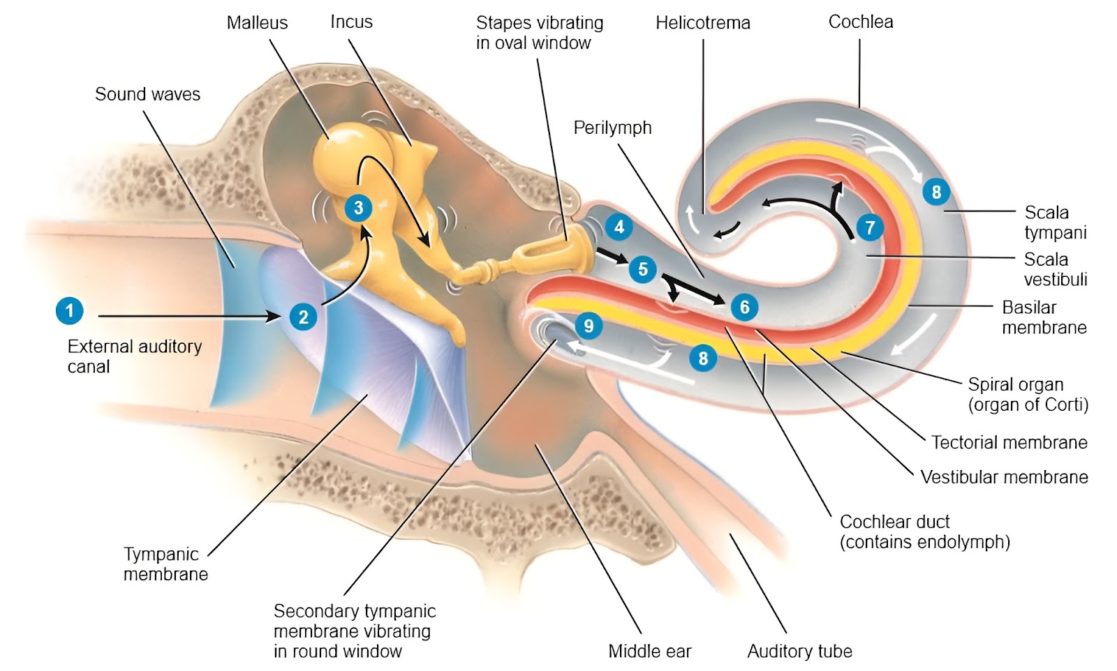
Perilymph in scala vestibuli catches or receives these vibrations and moves up and down. Ressner's membrane present in between scala vestibuli and scala media vibrates. Endolymph present in scala media moves up and down. Basilar membrane now receives these movements of endolymph and vibrates. Organ of corti, the hearing receptor, moves up and down. The hairs on the top of the hair cells of an organ of corti embedded in the tectorial membrane now tilt or bend. This mechanical stimulation i.e. bending of hairs produces action potential.
This action potential sums up and gives origin to nerve impulses, which are transmitted via the auditory pathway to the auditory center i.e. areas-41 in the temporal lobe of cerebral cortex. Neural signals from area-41 pass on to area-42, 22, 23 (auditory association areas) where interpretation of heard sounds is made.

Join the conversation