Anatomy and physiology of human eye
Anatomy of human eye :
The eyes are a pair of complex and highly developed sensory organs for vision. Sight or vision is extremely important for human survival. Large part of the cerebral cortex is devoted to processing visual information. The vision power of the eye is zero to infinity distance. More than half the sensory receptors in the human body are located in eyes (approximately 120 to 130 million photoreceptors). Each eye is spherical/rounded and called an eyeball. The diameter of an eyeball in an adult human is around 2.5 cm. Of its total surface area, only the anterior one-sixth is exposed, the remainder is recessed and protected by the orbit of the skull with a cushion of adipose tissue/fat around them.
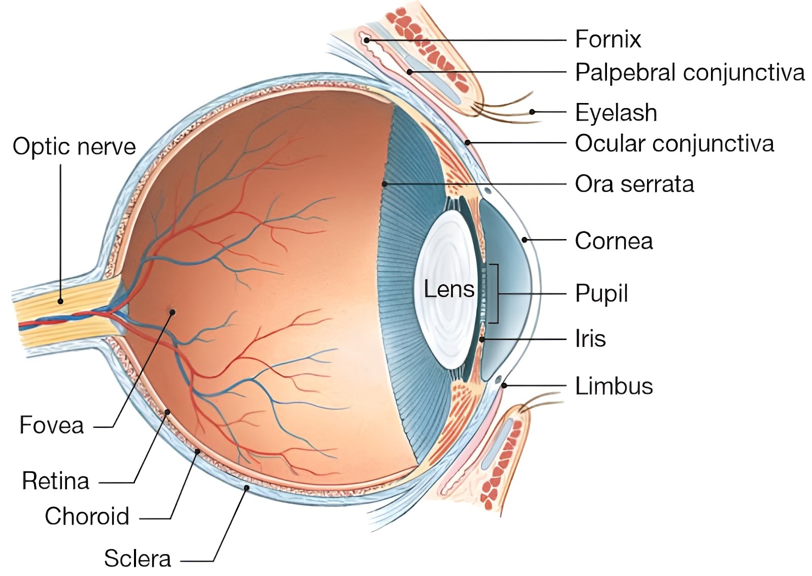
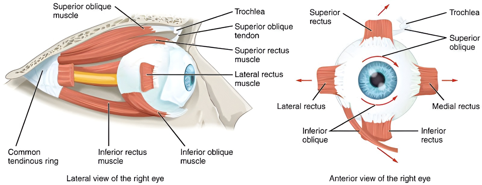
Movement of the eyeball within the orbit is controlled by 6 sets of muscles. These sets of muscles are controlled by cranial nerves, which are; Oculomotor (III), Trochlear (IV), and Abducens (VI) nerves.The eyes are protected by (or Accessory structures of the eye are); Bones, Eyebrows, Upper and lower eyelids, Eyelashes, Conjunctiva, and Lacrimal apparatus (i.e. include lacrimal/tear glands and lacrimal ducts).
Anatomically the wall of the eyeball consists of three layers/coats/tunica;
Outermost fibrous coat,
Middle vascular coat (Uvea), and
Innermost nervous coat (Ratina).
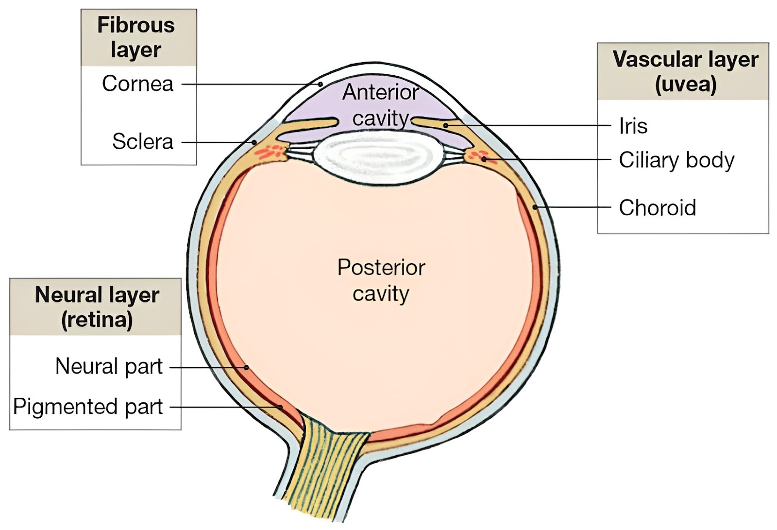

Fibrous coat :
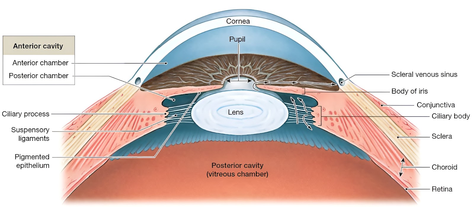
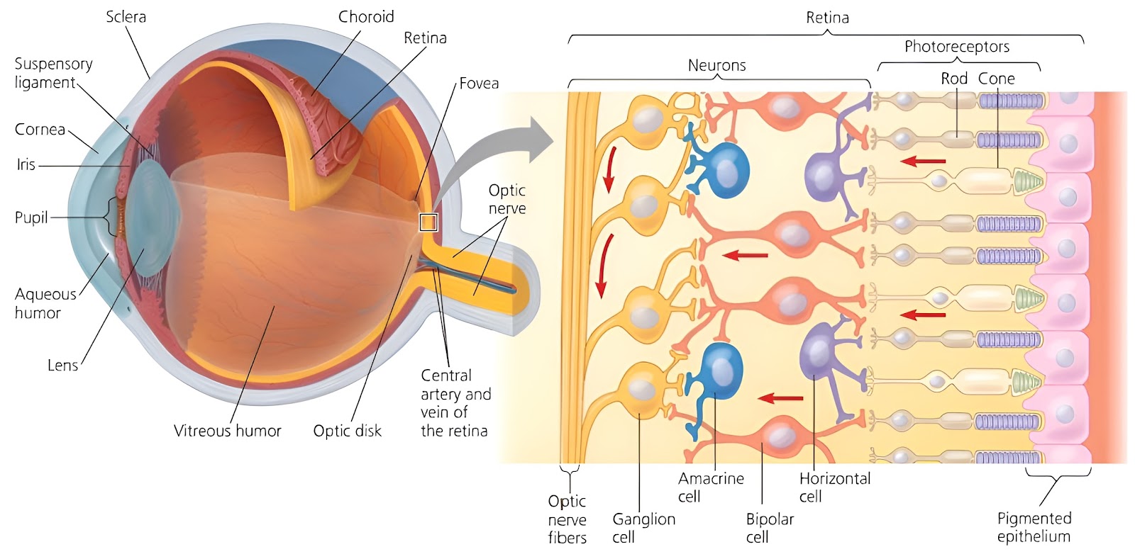
It is also known as Tunica fibrosa or Fibrous tunic or Corneoscleral coat. It is the complete superficial layer of eyeball, and consists of two parts; Posterior Sclera, and Anterior Cornea. The exposed part of sclera and the entire cornea are covered by a transparent membranous covering called conjunctiva. It provides protection and lubrication to the cornea. At the junction of the sclera and cornea is an opening known as the scleral venous sinus (Canal of Schlemm). A fluid called aqueous humour drains into this sinus.

Sclera or Scelrotic :
It covers the 5/6 posterior (or back) part of the eyeball. This layer is made of dense fibroelastic connective tissue with collagen fibres. It is the white part of the eye. And it covers the entire eyeball except the cornea. It protects and maintains the shape of the eyeball. And makes it more rigid. It provides attachment to the eyeball muscles, for eyeball movement. The sclera is provided with blood vessels, for oxygen and nutrition.
Cornea :
It covers the 1/6 anterior (or front) part of the eyeball. It is an extension of sclera, and it is a perfectly transparent, avascular layer. It is slightly bulged out for focussing light on the retina. The cornea allows free entry of light, and also provides maximum (about 75%) reflection to light. It gets nutrition from aqueous humour and also by lacrimal secretion. And it receives oxygen from the outside air. Comea has a rich nerve supply. It is very sensitive to touch & pain. Tears continuously flow over the cornea and keep it moist. The cornea was one of the first organs to be successfully transplanted.
Vascular coat (Uvea) :
It is also known as Tunica vasculosa or Vascular tunic or Uvea or Uveal tract. It is not a complete middle layer of eyeball, and consists of three parts; Choroid, Ciliary body, and Iris.

Choroid :
The choroid lies adjacent to the sclera. It contains numerous blood vessels that supply nutrients and oxygen to the other tissues, especially the retina. It also contains melanocytes that produce melanin pigment. Melanin causes this layer to appear dark brown in colour. Melanin absorbs stray light rays and converts the eyeball into a dark chamber.It prevents reflection and scattering of light within the eyeball. As a result, the image cast on the retina by the cornea and lens remains sharp and clear.
Ciliary body :
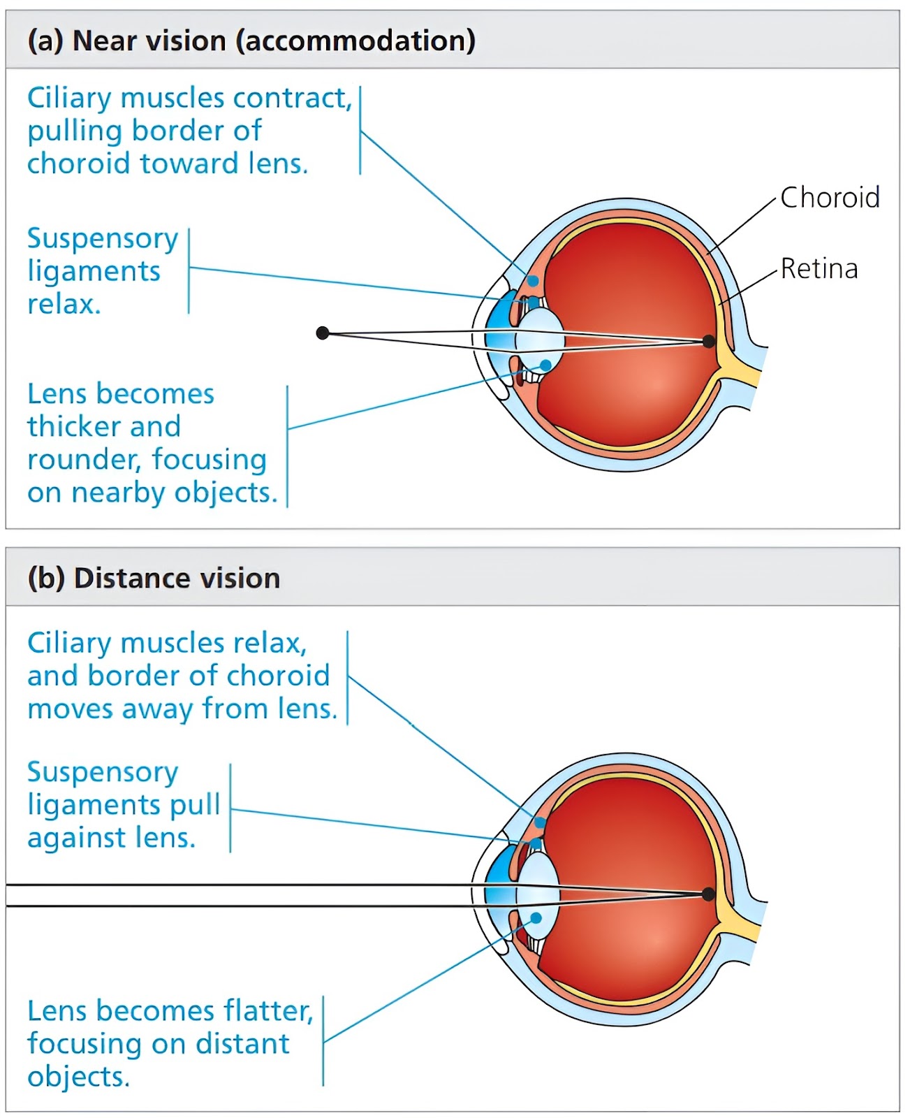
It is a thick, muscular, ring-like structure at the junction of choroid and iris. It appears dark brown in colour because it contains melanin-producing melanocytes. Peripherally it is smooth but centrally it has numerous finger-like processes called ciliary processes (70 to 80 in number). Ciliary processes are highly vascular and are sites for the formation of aqueous humour. Fine fibres called suspensory ligaments originate from ciliary processes and blend with the capsule of the lens, these hold the lens in position.
The ciliary muscle is a circular band of smooth muscle. Contraction or relaxation of the ciliary muscle changes the tightness of the zonular fibres or suspensory ligaments or zonule of zinn. It alters the shape of the lens, by adapting it for near or far vision. Nearby objects require more refraction to be focussed on the retina so the lens becomes more convex due to relaxation of suspensory ligaments (as a result of contraction of ciliary muscles), While faraway objects require less refraction to be focussed on retina so lens becomes less convex due to contraction of suspensory ligaments (as a result of dilation of ciliary muscles).
Iris :
It extends from the ciliary body across the eyeball in front of the lens. It separates the aqueous humour region into anterior and posterior chambers. It has an opening in the centre called the pupil. It consists of; Melanocytes, Circular smooth muscle fibres (or Constrictor pupillae), and Radial smooth muscle fibres (or Dilator pupillae). It is the colored portion of the eyeball. The amount of melanin pigment in the iris determines the eye colour. Eyes appear brown to black when the iris contains a large amount of melanin. Eyes appear blue when its melanin concentration is very low. Eyes appear green when its melanin concentration is moderate. Eye colour depends upon; Thickness of Iris, Amount of melanin, and Distribution of melanin. The pattern of iris is unique to every individual.
Nervous coat (Retina) :
It is also known as Tunica nervosa or Retina. It has the innermost, delicate, avascular light sensitive layer. It has two regions;
Outer pigmented non-sensory part, and
Inner nervous sensory part.
Pigmented non-sensory part :
It is single layered, lining the iris and ciliary body. It has cuboidal epithelial cells. It engulfs discs of the outer segment of rods and cones cells.
Nervous sensory part :
It is triple layered, lining the choroid. These three layers are; Outer photosensitive layer of rod and cone cells (or first order neurons), Middle layer of bipolar nerve cells (or second order neurons), and Inner layer of ganglion cells (or third order neurons). The nerve fibres from the basal end of the ganglion cells collectively form the optic nerve. Optic nerve and also arteries and veins pass through the optic disc (or Blind spot). The external surface of the retina is in direct contact with the choroid and its internal surface with the vitreous humour.
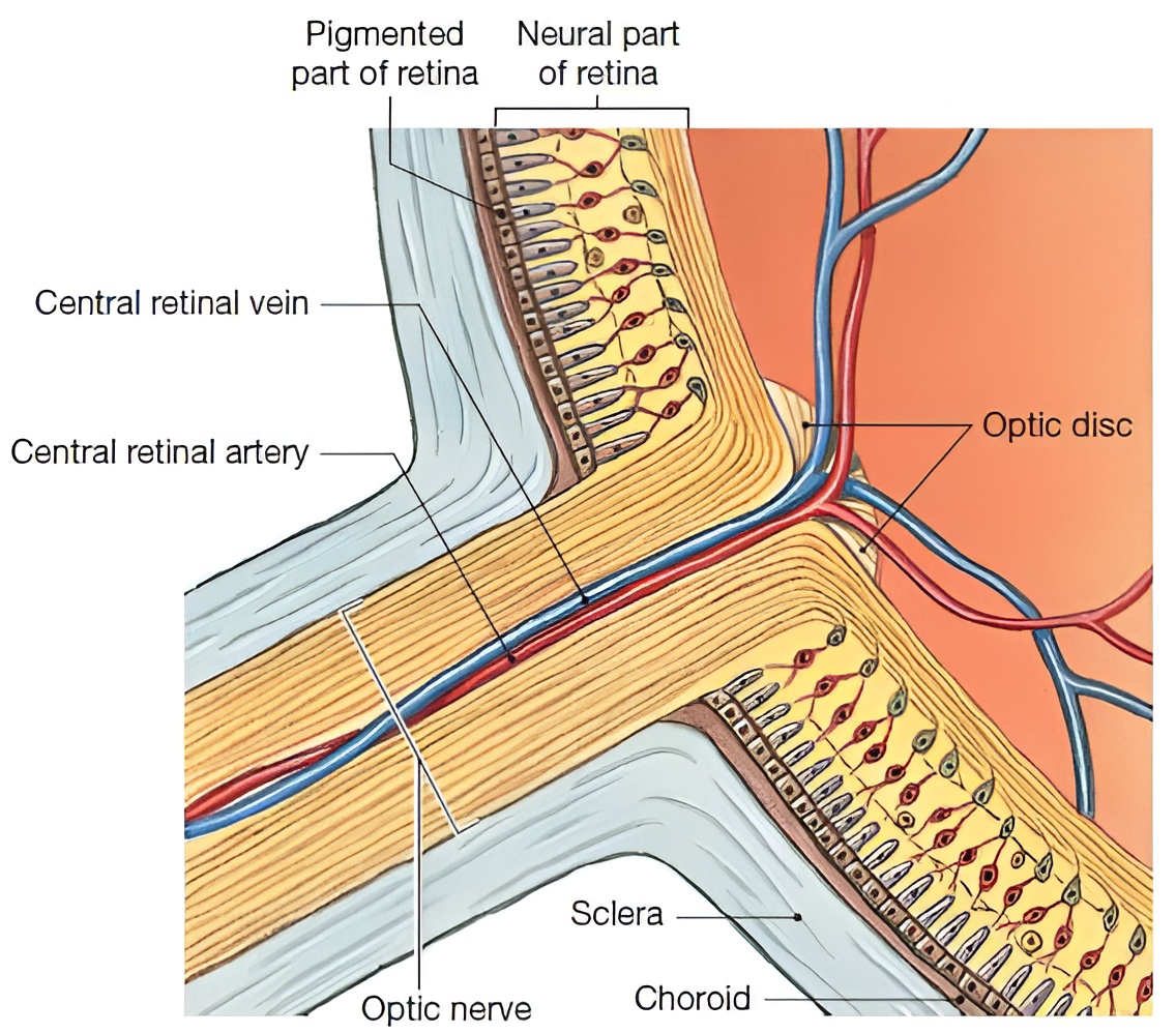
Blind spot (or Macula lutea) and Yellow spot (or Optic disk or Dead spot or Scotoma);
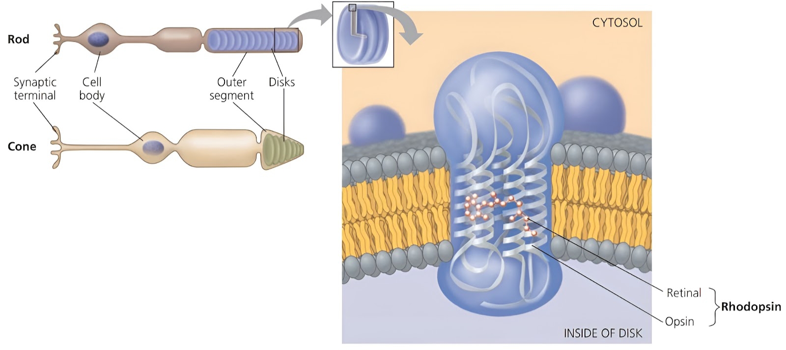
First order neurons or Photoreceptors :
These are modified bipolar neurons. These are embedded in the pigment layer of the retina. Structurally there are two segments of photoreceptors; Outer segment, and Inner segment. The outer segment of rod has thousands of flattened discs, while the inner segment has the main metabolic machinery like mitochondria, and Golgi body, etc. The synaptic terminal forms synapse with dendrites of bipolar neurons. The plasma membrane of the disc has light sensitive proteins termed as photopigments molecules.

Few comparative information about rods and cones of human eye;

Pupil :
Pupil is the hole (or aperture) in the centre of the iris. Pupil appears black because, as you look through the lens, you see the heavily pigmented back of the eye. Its diameter varies from 2mm to 8mm (normally it is 3 to 4 mm). Pupils are larger in females as compared to males. It allows free entry of light. It constricts in bright light and dilates in dim light.
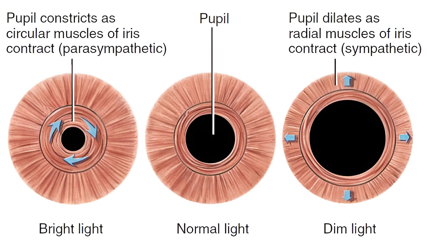
Constriction of the pupil is called Miosis, While Dilation of pupil is called Mydriasis. After death the pupil dilates and does not respond to light. Drugs which cause miosis are miotics, Example; Acetylcholine. While drugs which cause mydriasis are mydriatics, Example; Atropine. Parasympathetic nervous system is responsible for miosis, while the sympathetic nervous system is responsible for mydriasis.
Lens :
It is a transparent, elastic, biconvex, living structure which is capable of changing its refractive power and this property of lens is called the Accommodation power. It is suspended in the eyeball by the suspensory ligaments. The lens and suspensory ligaments divide the cavity of the eyeball into; Small anterior aqueous chamber, and Large posterior vitreous chamber. Anterior chamber is filled with a clear watery fluid called Aqueous humour, while the posterior chamber is filled with a jelly-like substance called Vitreous humour.
Lens is made up of thousands of layers of cells which have crystallin proteins. Lens cells lose their nucleus and other cell organelles during development to become perfectly transparent. It gets nutrition from aqueous humour (present on front as well as back of lens). Lens continues to grow throughout life. With advancing area there is decrease in elasticity of lens called Presbyopia.
Lacrimal apparatus :
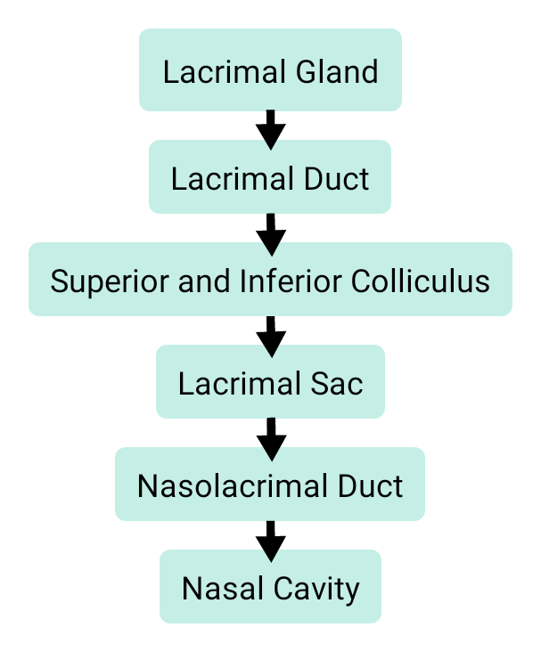
The lacrimal apparatus of each eye consists of; Lacrimal gland, Lacrimal ducts, Superior and inferior lacrimal canaliculi/canals, Lacrimal sac, and Nasolacrimal duct. The lacrimal gland is situated in the orbit on the superolateral surface of the eyeball. The lacrimal gland secretes tears. The composition of tears is; Water, Salts, and Bactericidal protein called Lysozyme. The flow of tears in the lacrimal apparatus is as follow;

Accommodation power of lens :
The ciliary muscle is a circular band of smooth muscle. Contraction or relaxation of the ciliary muscle changes the tightness of the Zonular fibres or Suspensory ligaments or zonule of zinn. It alters the shape of the lens, by adapting it for near or far vision. Nearby objects require more refraction to be focussed on the retina so the lens becomes more convex due to relaxation of suspensory ligaments (as a result of contraction of ciliary muscles), while faraway objects require less refraction to be focussed on retina so lens becomes less convex due to contraction of suspensory ligaments (as a result of dilation of ciliary muscles).
Extraocular muscles of eye :
Movement of the eyeball within the orbit is controlled by six sets of muscles. These muscles are called extraocular muscles, and tendons of these muscles are attached to sclera. There are four Rectus Muscles and two Oblique muscles. These sets of muscles are controlled by cranial nerves, which are; Oculomotor (III), Trochlear (IV), and Abducens (VI).
Physiology of vision :
The light rays from the object pass through; Conjunctiva, Cornea, Anterior chamber, Pupil, Lens, Posterior chamber, and then focused on the photoreceptors (include rods and cones) and changes cis-retinal to trans-retinal. During this change, cGMP is broken down to GMP (sodium ions no longer flow into the cell). Lack of sodium ions causes hyper-polarisation of the cell and this stops the continual release of glutamic acid neurotransmitter. This lack of inhibitory glutamic acid stimulates the bipolar cells. The bipolar cells stimulate the ganglionic cells to produce an action potential.
Action potential passes via; Optic nerve, Optic chiasma, Optic tract, and relay in lateral geniculate body (a nucleus of thalamus). Fibres go to superior colliculus and pretectal area; for example, for pupillary reflexes (i.e. light and accommodation reflexes) and then pass through Optic radiation. Fibres terminate in primary visual area-17 of the occipital cortex, where the true image of the object is formed. Neural signals pass to visual association area-18 for recognition and interpresentation of visualised objects.

Glaucoma :
It is also known as Kaala Motia, it is a painful condition. Due to blockage of the canal of schlemm (present at junction of cornea and sclera). There is an increase in intraocular pressure. Vision lost due to glaucoma cannot be regained back.
Join the conversation